DeePathology™ STUDIO web

Bringing the power of AI to pathology teams!

  • Powerful AI algorithms

    The DeePathology™ STUDIO web is an interactive Do it Yourself platform for AI in pathology. Researchers and pathologists can create powerful AI solutions for histological image analysis and use these algorithms to create powerful analytics on new pathology slides. Create proof of concepts (PoC) for most problems - in 5 minutes, no computer coding required!

  • Lightning-fast annotation

    Do you want to annotate hundreds of thousands of cells in just a few days? With the DeePathology™ STUDIO web, your team can achieve this goal using our innovative “Deep Interactive Online Active Learning”.

  • Large-scale operations

    Identify hundreds of millions of cells in your slides by deploying your algorithms in the cloud for large-scale quantification projects.

  • Custom seamless integration

    As a server-based platform, the DeePathology™ STUDIO web can be seamlessly integrated into any professional workflow, for example to communicate with your image management software.

  • Multiple user projects

    Unlimited users can work on the same project simultaneously to use precious resources most effectively. Sharing direct links to slide locations at any time encourages collaboration to create the best ground truth possible in the least amount of time.

  • High data security

    The DeePathology™ STUDIO web is easy to use and fun while keeping your data private. It is installed on a server on your premises, so you retain full control over your sensitive data without having to send it to a remote server.

  • Versatile and flexible

    Detect and classify cells and other objects, segment tissue regions and classify image tiles - all in one product. Combine modes to create multi-layered solutions, for example precise cell quantification within specific tissue regions.

Experience the DeePathology™ STUDIO web in action!

DeePathology™ STUDIO web

3 Videos
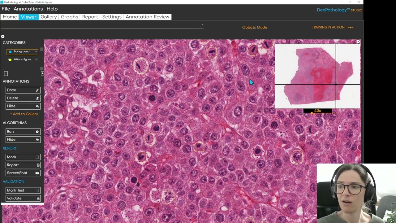
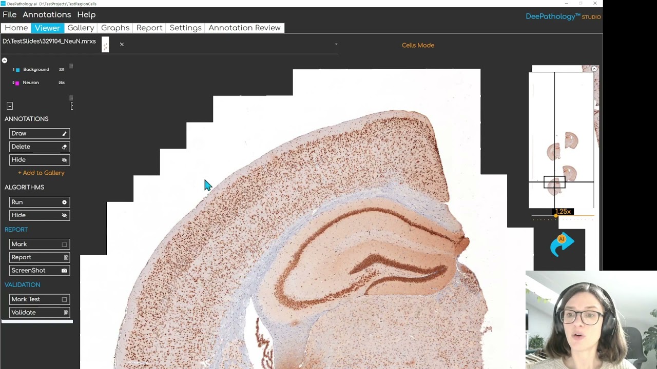
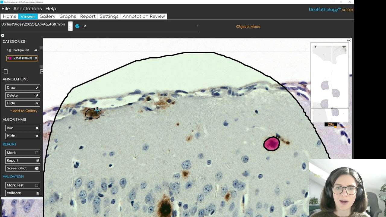
The DeePathology™ STUDIO web is a revolution for AI-powered pathology research!
Jacob Gildenblat
Chief Technology Officer and Co-Founder

The ping pong Workflow:

Pathologist vs. DeePathology™ STUDIO web

Annotate

Annotate examples from different categories, using the fast annotation modes in the DeePathology™ STUDIO web.

Interact

Interact with the DeePathology™ STUDIO web by running it on field of views, correcting mistakes and quickly adding new training data using the powerful Interactive Online Active Learning engine.

Analyze

Once you are ready to go, you can apply your algorithm to new slides and create powerful analytic reports.

One AI Platform -

Countless Pathology Use Cases

H&E
  • Quantify tumor infiltrating lymphocytes (TILs)
  • Segment tissue in tumor and healthy tissue
  • Detect glandular structures, such as glomeruli, acini, …
  • Quantify mitotic figures
  • Segment the growth plate region in bone sections
  • Detect red blood cells
  • Segment lung tissue and quantify cells in wall regions
  • Detect granulated and degranulated cells in skin cancer
  • Detect regions with high lymphocyte infiltration in tumor sections
IHC
  • Quantify Ki67 positive cells and calculate the stain ratio
  • Segment positive and negative regions, for example in FoxP3, CD7 and other immunostains
  • Detect different types of beta-amyloid plaques in Alzheimer’s disease research
  • Quantify glia cells in neuroscience research, including astrocytes and microglia
Other stains
  • Quantify cells of interest in in-situ hybridization (ISH) stains
  • Detect Helicobacter pylori in H&E or Giemsa stained slides
  • Detect regions infected with Mycobacterium tuberculosis in Ziehl-Neelsen stained slides for veterinary TB surveillance
A huge time saver for CROs and pharma companies engaged in large-scale projects with big teams in multiple locations.
Chen Sagiv, PhD
Co-CEO and Co-Founder

Four modes for unlimited possibilities

DeePathology™ STUDIO Web Objects Mode

  • Object segmentation

    Create algorithms that detect the boundaries of objects and classify them into unlimited categories.

  • Optimized for small data

    Annotate as few as four to six examples from each category to get started. Then run the algorithm and correct mistakes - your proof of concept solution is ready in minutes.

  • Annotation magic

    The DeePathology™ STUDIO web also finds objects for you and displays them in the gallery. The gallery leverages an extremely powerful Active Learning Engine, suggesting you the most important examples to annotate. All you have to do is to set their category and you will obtain a high-quality balanced dataset in no time.

DeePathology™ STUDIO Web Cells Mode

  • Cell detection and classification

    Get accurate detections in challenging real life scenarios by detecting cells with our brand new H&E cell detector. Classify a small number of examples, then detect millions of them in new slides. The cells mode also includes an IHC-focused detection algorithm and a re-trainable detection algorithm for custom needs.

  • Powerful metrics

    Monitor the performance of your algorithm in real-time - you automatically obtain basic validation analytics for each epoch.

  • Annotation magic

    Using the gallery, the DeePathology™ STUDIO web makes sure that you are annotating the most important cells. With the AI mode, you can annotate thousands of cells in just a few minutes. And it is not just the quantity of cells - these are high quality images that will make your dataset diverse, and improve your algorithms the most. All you have to do is sit back, drink some coffee, and correct mistakes from time to time in the gallery.

DeePathology™ STUDIO Web Regions Mode

  • Region segmentation

    Create region segmentation algorithms for detecting regions of different types, for example different tissues or tumor regions vs. healthy tissue.

  • Multilayered analyses

    You can then detect these region types in new slides, and combine the algorithm with other DeePathology™ STUDIO modes to count different types of cells and objects within the different regions.

We need images from tiles mode. 🙂

DeePathology™ STUDIO Web Tiles Mode

  • Tile classification

    Quickly annotate tiles (small squares) with single clicks in the viewer.

  • Optimized for speed

    Use the gallery to annotate thousands of tiles from different slides in just minutes. Annotating tiles is simple, fast, and lets you create powerful solutions in record time.

Frequently Asked Questions:

DeePathology™ STUDIO web is installed on a server on your premises. This can be a Windows or Linux server.

If you prefer a solution that can be installed locally on your computer, the DeePathology™ STUDIO desktop may be the right choice for you.

We support all major scanner WSI formats (.svs, .ndpi, .tiff, .bif, .mrxs and more), and also regular images (.png, .jpeg, .bmp and so on).

Please contact us for further information about licencing conditions and prices.

Get In Touch With Us Today!

Request your free personal demonstration and learn how the DeePathology™ STUDIO web can be integrated into your workflow.

We use cookies to provide the services and features offered on our website, and to improve our user experience. Visit our Privacy Policy to learn more.

LOGIN