Ki-67 has gained relevance in oncology as a marker of proliferating cells. Particularly breast cancer patients benefit from this additional diagnostic marker and subsequent therapeutic decisions. However, breast cancer is not the only cancer type where Ki-67 immunohistochemistry is of interest.
Dr. Kirsten Utpatel, pathologist at the University of Regensburg, Germany, has developed an algorithm for Ki-67 quantification in liver tissue. The algorithm was trained in the DeePathology™ STUDIO to detect two classes of hepatocytes based on their Ki-67 expression while ignoring other types of cells, e.g. lymphocytes and Kupffer cells.

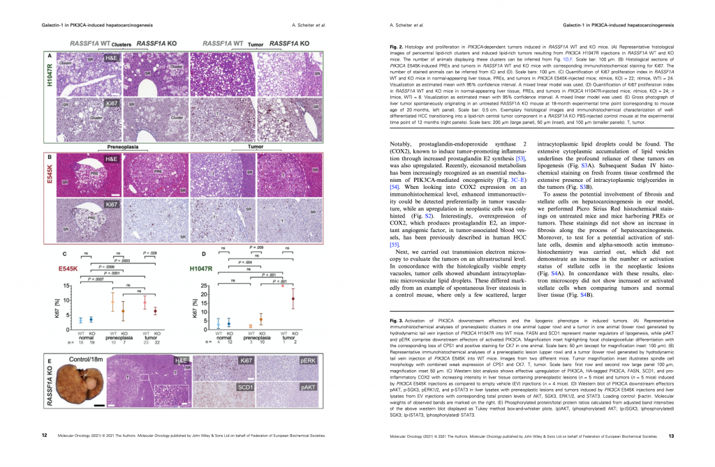
In their recent publication, Dr. Utpatel and colleagues used the algorithm to determine the ratio of Ki-67-positive to Ki-67-negative hepatocytes in tumor, preneoplastic regions and surrounding tissue (Scheiter et al., 2021). The study aimed to investigate the implications of two major signaling pathways, the mTOR pathway and the RAS/MAPK pathway, on the formation of experimental hepatocellular carcinomas and identified potential novel treatment options. Read the full, open-access article published in “Molecular Oncology”.
Dr. Utpatel has previously developed an algorithm for prostate cancer classification, which she presents in a webinar about her experience with the DeePathology™ STUDIO. Have a look at this and other webinars!

